pelvic ultrasound scan

Ultrasound Scan for Womens Health

ultrasound scan pelvis and abdominal

Polycystic ovarian syndrome, uterine fibroids, ovarian cysts, pelvic inflammatory disease, pelvic organ prolapse — all of these conditions have one thing in common. They’re diagnosed using pelvic ultrasound technology.

Developed by a team of Scottish researchers over 50 years ago in the aftermath of World War II, this technology has come a long way since then and is, quite literally, saving lives the world over.

Learn more about what to expect from a Wellwoman scan and why ultrasound technology is so important for women’s health.

Pelvic Ultrasound: A Closer Look

Also referred to as sonography, ultrasounds are both popular and beneficial because of how efficient and accurate they are. They’re also non-invasive, which means that there’s no downtime associated with this test.

Ultrasound technology uses very high frequency sound waves to capture detailed images of the internal structure of the body. The end result is a sonograph. It appears as white pixels against a black background, detailing internal organs or areas of soft tissue.

In the case of a pelvic ultrasound, this includes organs such as the bladder, ovaries, fallopian tubes, the lining of the uterus, vagina, and rectum. A pelvic ultrasound may also encompass areas of the lower abdomen.

Ultrasound scanning is the best way for a doctor to see and assess what’s happening inside the body, without having to cut you open.

How is an ultrasound different from an x-ray, you might wonder?

While the overall premise is the same, an ultrasound can provide more detailed imagery in some cases, and does not expose you to radiation.

A pelvic ultrasound is somewhat of an umbrella term that encompasses several different types of sonography. Let’s take a look at the different types of ultrasound beneficial to women:

private ultrasound scan

An Abdominal Ultrasound

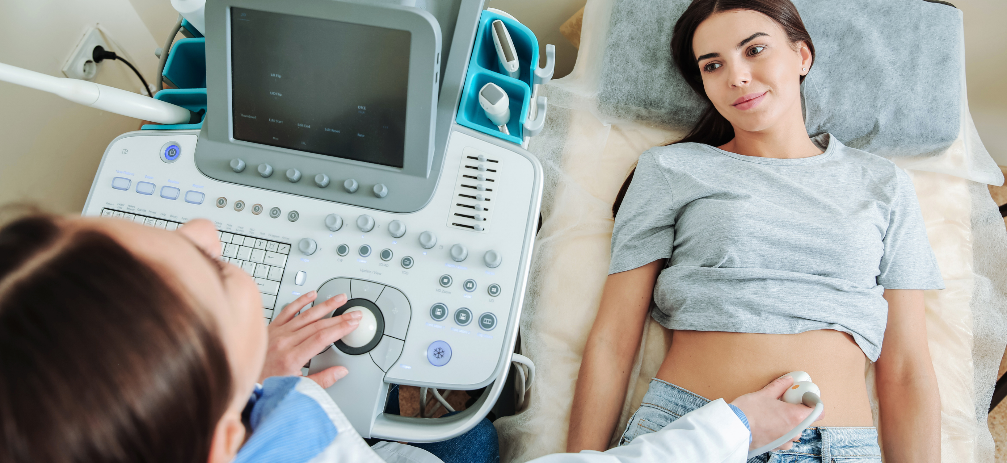
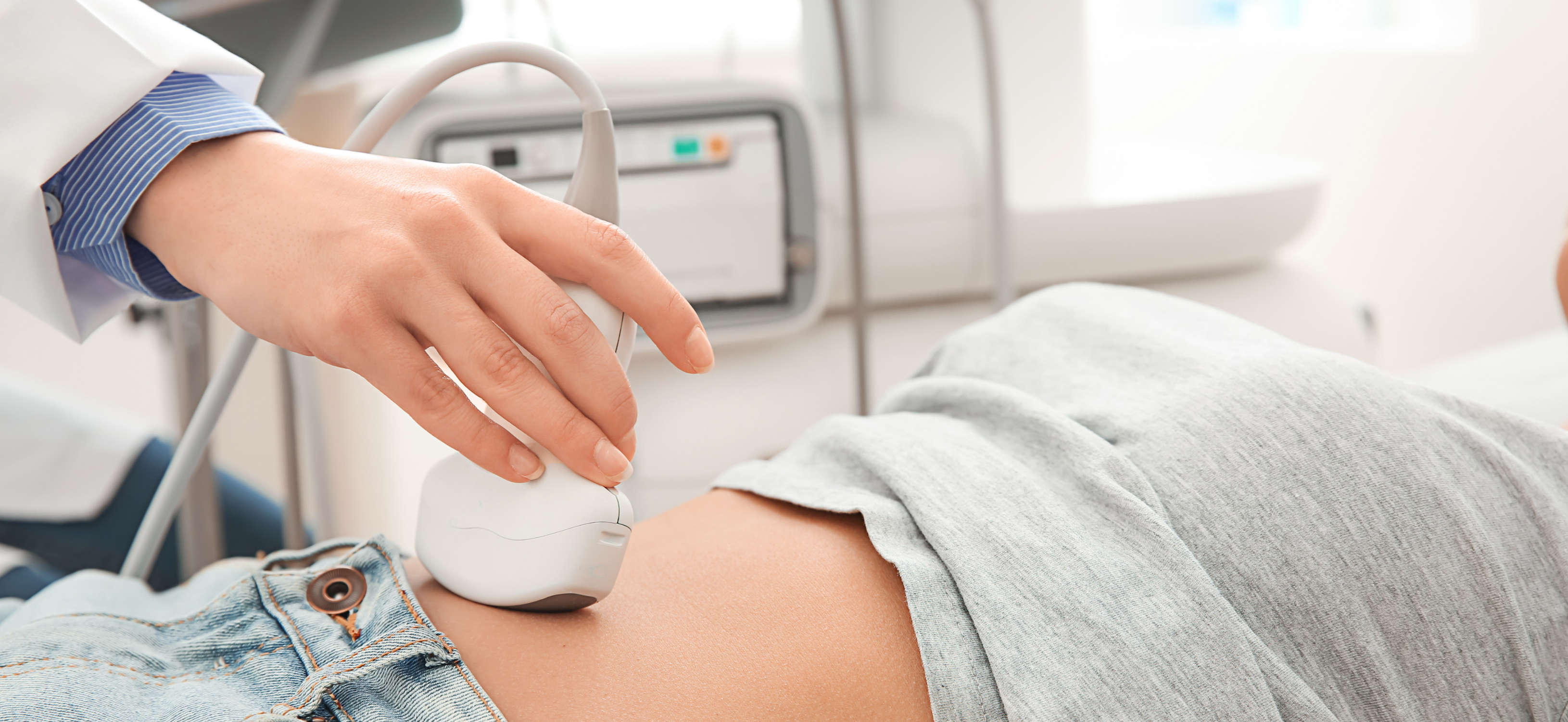
The purpose of this test is to analyse organs inside the belly, specifically, the lower part of the belly.

During the ultrasound, a sonographer applies warm gel to the lower abdomen area. They then use a tool known as a transducer, pressing it onto the skin to see the organs and soft tissue underneath.

The gel helps the transducer move easily over your skin during the scan. While you could have an x-ray of your abdomen, it does not offer as much detail as ultrasound, especially if you have renal stones or gallstones.

If you have abdominal pain, have experienced trauma to the abdomen, or even have unexplained uterine pain, an abdominal scan is useful.

A Transvaginal Ultrasound

This type of scan is incredibly beneficial in women because it examines several different reproductive organs at once, from inside the vagina.

For example, a sonographer can examine the vagina, uterine lining, fallopian tubes, pelvic cavity, bladder, and ovaries in just one scan.

During this ultrasound, a sonographer inserts a transducer into the vagina. The transducer is covered with a warm gel to assist with lubrication. They then move the transducer around gently to obtain imagery of different reproductive organs.

When Is a Pelvic Ultrasound Necessary?

So, when is it a good idea to visit your doctor and when might they recommend any of these pelvic ultrasounds?

Some of the most common troublesome symptoms include:

  • Painful sexual intercourse
  • Infertility — issues trying to fall pregnant
  • Painful urination, i.e. a burning sensation when urinating
  • Unexplained pelvic or abdominal pain
  • Unexplained or sudden swelling in your abdomen
  • Trauma to the abdomen or pelvic area
  • Irregular or exceptionally painful menstruation and PMS symptoms
  • Menstruation after menopause
  • Prolapse/incontinence

Thanks to the innovative technology of ultrasound, it can diagnose a myriad of different conditions. From hernias to kidney stones, ovarian cysts, polycystic ovarian syndrome (PCOS), ectopic pregnancy, and more.

Not only is ultrasound helpful in diagnosing these conditions, but it’s also used for important procedures such as biopsies and IUD placement in the uterus.

Preparing for an Ultrasound: What To Expect

Different types of pelvic ultrasounds require different levels of preparation. For some, you don’t need to prepare much at all.

When it comes to pelvic ultrasounds of the reproductive organs, you’ll need to arrive for your scan with a full bladder. Begin drinking about one litre of water an hour before your scan so that it’s nice and full when you arrive.

For an ultrasound of the abdomen, you might need to fast for a few hours before your scan. This means cutting out food or drinks (except water) for at least 6 hours before the test.

Bear in mind that the doctor who refers you for an ultrasound will brief you on how to prepare before the scan. You might also receive instructions via email or telephone a few days before.

Is It Painful?

Ultrasound scans are completely harmless and do not cause any pain. This is just one of the reasons they’re so widely used today. For the most part, they’re performed over the skin.

The most discomfort you might feel is a feeling of pressure against a full bladder.

Transvaginal or rectal scans are a little different. While they’re by no means painful, they are uncomfortable for some women. The important thing is to relax and breathe, and trust that your sonographer knows exactly what they’re doing.

Looking For Private Ultrasound Scanning?

If you need a pelvic ultrasound and prefer the idea of a private clinic, then our team at Bothwell Medical Rooms & Core Clinic are your Glasgow and Lanarkshire go-to for a Wellwoman scan.

We offer private clinics in both Bothwell and Hamilton for all your diagnostic needs. Get in touch with our team for efficient service that fits in with your schedule.

book now

ultrasound scan pelvis and abdominal

Pelvic Ultrasound

What is a pelvic ultrasound?

A pelvic ultrasound is a noninvasive diagnostic exam that produces images that are used to assess organs and structures within the female pelvis. A pelvic ultrasound allows quick visualization of the female pelvic organs and structures including the uterus, cervix, vagina, fallopian tubes, and ovaries.

Ultrasound uses a transducer that sends out ultrasound waves at a frequency too high to be heard. The ultrasound transducer is placed on the skin, and the ultrasound waves move through the body to the organs and structures within. The sound waves bounce off the organs like an echo and return to the transducer. The transducer processes the reflected waves, which are then converted by a computer into an image of the organs or tissues being examined.

The sound waves travel at different speeds depending on the type of tissue encountered – fastest through bone tissue and slowest through the air. The speed at which the sound waves are returned to the transducer, as well as how much of the sound wave returns, is translated by the transducer as different types of tissue.

An ultrasound gel is placed on the transducer and the skin to allow for smooth movement of the transducer over the skin and to eliminate air between the skin and the transducer for the best sound conduction.

Another type of ultrasound is Doppler ultrasound, sometimes called a duplex study, used to show the speed and direction of blood flow in certain pelvic organs. Unlike a standard ultrasound, some sound waves during the Doppler exam are audible.

Pelvic ultrasound may be performed using one or both of 2 methods:

  • Transabdominal (through the abdomen). A transducer is placed on the abdomen using the conductive gel
  • Transvaginal (through the vagina). A long, thin transducer is covered with the conducting gel and a plastic/latex sheath and is inserted into the vagina

The type of ultrasound procedure performed depends on the reason for the ultrasound. Only one method may be used, or both methods may be needed to provide the information needed for diagnosis or treatment.

Other related procedures that may be used to evaluate problems of the pelvis include hysteroscopy, colposcopy, and laparoscopy.

../../GetImage.aspx?ImageId=161348

What are female pelvic organs?

The organs and structures of the female pelvis are:

  • Endometrium. The lining of the uterus
  • Uterus (also known as the womb). The uterus is a hollow, pear-shaped organ located in a woman’s lower abdomen, between the bladder and the rectum. It sheds its lining each month during menstruation unless a fertilized egg (ovum) becomes implanted and pregnancy follows.
  • Ovaries. Two female reproductive organs are located in the pelvis in which egg cells (ova) develop and are stored and where the female sex hormones estrogen and progesterone are produced.
  • Cervix. The lower, narrow part of the uterus is located between the bladder and the rectum, forming a canal that opens into the vagina, which leads to the outside of the body.
  • Vagina (also known as the birth canal). The passageway through which fluid passes out of the body during menstrual periods. The vagina connects the cervix and the vulva (the external genitalia).
  • Vulva. The external portion of the female genital organs

What are the reasons for a pelvic ultrasound?

Pelvic ultrasound may be used for the measurement and evaluation of female pelvic organs. Ultrasound assessment of the pelvis may include, but is not limited to, the following:

  • Size, shape, and position of the uterus and ovaries
  • Thickness, echogenicity (darkness or lightness of the image related to the density of the tissue), and presence of fluids or masses in the endometrium, myometrium (uterine muscle tissue), fallopian tubes, or in or near the bladder
  • Length and thickness of the cervix
  • Changes in bladder shape
  • Blood flow through pelvic organs


Pelvic Ultrasound – Bothwell Medical Rooms

ultrasound scan

Pelvic ultrasound can provide much information about the size, location, and structure of pelvic masses, but cannot provide a definite diagnosis of cancer or specific disease. A pelvic ultrasound may be used to diagnose and assist in the treatment of the following conditions:

  • Abnormalities in the anatomic structure of the uterus, including endometrial conditions
  • Fibroid tumors (benign growths), masses, cysts, and other types of tumors within the pelvis
  • Presence and position of an intrauterine contraceptive device (IUD)
  • Pelvic inflammatory disease (PID) and other types of inflammation or infection
  • Postmenopausal bleeding
  • Monitoring of ovarian follicle size for infertility evaluation
  • Aspiration of follicle fluid and eggs from ovaries for in vitro fertilization
  • Ectopic pregnancy (pregnancy occurring outside of the uterus, usually in the fallopian tube)
  • Monitoring fetal development during pregnancy
  • Assessing certain fetal conditions

There may be other reasons for your doctor to recommend a pelvic ultrasound.

What are the risks of a pelvic ultrasound?

There is no radiation used and generally no discomfort from the application of the ultrasound transducer to the skin during a transabdominal ultrasound. You may experience slight discomfort with the insertion of the transvaginal transducer into the vagina.

Transvaginal ultrasound requires covering the ultrasound transducer in a plastic or latex sheath, which may cause a reaction in patients with a latex allergy.

During a transabdominal ultrasound, you may experience discomfort from having a full bladder or lying on the examination table.

If a transabdominal ultrasound is needed quickly, a urinary catheter may be inserted to fill the bladder.

There may be risks depending on your specific medical condition. Be sure to discuss any concerns with your doctor prior to the procedure.

Certain factors or conditions may interfere with the results of the test. These include, but are not limited to, the following:

  • Severe obesity
  • Barium within the intestines from a recent barium procedure
  • Intestinal gas
  • Inadequate filling of the bladder (with transabdominal ultrasound). A full bladder helps move the uterus up and moves the bowel away for better imaging.

How do I prepare for a pelvic ultrasound?

EAT/DRINK: Drink a minimum of 1-2 litres of clear fluid at least one hour before your appointment. Do not empty your bladder until after the exam.

Generally, no fasting or sedation is required for a pelvic ultrasound, unless the ultrasound is part of another procedure that requires anesthesia.

For a transvaginal ultrasound, you should empty your bladder right before the procedure.

Your doctor will explain the procedure to you and offer you the opportunity to ask any questions that you might have about the procedure.

Based on your medical condition, your doctor may request other specific preparation.

book now

What happens during a pelvic ultrasound?

A pelvic ultrasound may be performed in your doctor’s office, on an outpatient basis, or as part of your stay in a hospital. Procedures may vary depending on your condition and your hospital’s practices.

Generally, a pelvic ultrasound follows this process:

Illustration of a transabdominal ultrasound procedure" height=

For a transabdominal ultrasound

  1. You will be asked to remove any clothing, jewelry, or other objects that may interfere with the scan.
  2. If asked to remove clothing, you will be given a gown to wear.
  3. You will lie on your back on an examination table.
  4. A gel-like substance will be applied to your abdomen.
  5. The transducer will be pressed against the skin and moved around over the area being studied.
  6. If blood flow is being assessed, you may hear a “whoosh, whoosh” sound when the Doppler probe is used.
  7. Images of structures will be displayed on the computer screen. Images will be recorded on various media for the health care record.
  8. Once the procedure has been completed, the gel will be removed.
  9. You may empty your bladder when the procedure is completed.
Illustration of transvaginal ultrasound procedure

For a transvaginal ultrasound

  1. You will be asked to remove any clothing, jewelry, or other objects that may interfere with the scan.
  2. If asked to remove clothing, you will be given a gown to wear.
  3. You will lie on an examination table, with your feet and legs supported for a pelvic examination.
  4. A long, thin transvaginal transducer will be covered with a plastic or latex sheath and lubricated. The tip of the transducer will be inserted into your vagina. This may be slightly uncomfortable.
  5. The transducer will be gently turned and angled to bring the areas for study into focus. You may feel mild pressure as the transducer is moved.
  6. If blood flow is being assessed, you may hear a “whoosh, whoosh” sound when the Doppler probe is used.
  7. Images of organs and structures will be displayed on the computer screen. Images may be recorded on various media for the health care record.
  8. Once the procedure has been completed, the transducer will be removed.

What happens after a pelvic ultrasound?

There is no special type of care required after a pelvic ultrasound. You may resume your normal diet and activity unless your doctor advises you differently.

There are no confirmed adverse biological effects on patients or instrument operators caused by exposures to ultrasound at the intensity levels used in diagnostic ultrasound.

Your doctor may give you additional or alternate instructions after the procedure, depending on your particular situation.

For further details or to book a pelvic ultrasound please contact the team on 01698 854474 or email info@bothwellmedicalrooms.co.uk

bothwell medical rooms

Wellwoman Pelvic Scan

Wellwoman Pelvic Scan – Diagnostic Ultrasound

A wellwoman pelvic ultrasound scan is a medical procedure. It captures live images from inside your body using high-frequency sound waves. It’s also known as sonography.

What are the benefits of choosing a private ultrasound?

Going private not only gives you the flexibility to work around your schedule. It also gives you faster diagnostic and earlier information. There are many reasons why going private may be the best solution for you. With the recent pandemic and many organisations dealing with months of backlog, the long wait times can often lead to a delay in diagnostics. This often leads to an increase in anxiety for many people.

Why Choose Us?

We offer the most accessible and affordable diagnostic imaging in Lanarkshire. Access to quick and effective diagnosis should be possible for everyone. We are a leading service within our field, recognisable for the top-quality service we provide. An efficient, compassionate team of professionals with a client 1st approach to healthcare. We specialise in diagnostic ultrasounds. This means all our exams are performed and reported by our expert Consultant Sonographers and Radiographers.

Pelvic Scan – Well Woman


This gynecological scan is available to women of pre-menopausal age.

We will perform a detailed scan to look at the size, shape, and condition of the:

  • uterus
  • the lining of the womb
  • both ovaries
  • pelvic area

Common examples for having this ultrasound scan are the detection or exclusion of:

  • fibroids
  • ovarian cysts
  • polycystic ovaries
  • pelvic inflammatory disease (PID)
  • IUCD(coil) location

We will provide a Sonographer report within 24 hours of examination taking place with a Medical Follow Up Recommendation if necessary.

Pre-scan preparation

May include transvaginal/internal scan with consent.

Need to expose lower part of the abdomen so ideally wear 2 piece garments i.e trousers or skirt and top.

Drink 0.75 Ltr (1.5 pints) clear fluid approx 1 hour before the appointment time. Milk and fizzy drinks are not recommended.

Both transabdominal and transvaginal scans will be performed for optimum clarity and results. Please do not empty your bladder prior to examination.

*if you choose not to have a transvaginal scan we may be able to complete the scan trans abdominally however please note that results may not be as accurate.

Contact the team today on 01698 854474 or email info@bothwellmedicalrooms.co.uk


Ultrasound Scan Costs